Obstetric or Pregnancy ultrasound examinations can be performed in all three trimesters during a pregnancy to assess overall growth and development of the foetus.
Specific examinations include
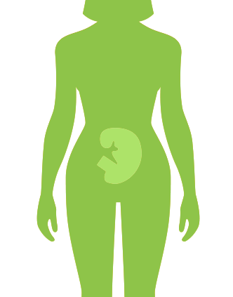
| Early Pregnancy Ultrasound Scans This ultrasound examination is performed mainly at 5 -12 weeks gestation.
The scan can be performed trans-abdominal, with the ultrasound probe gliding gently across the surface of your abdomen or endovaginal to allow better visualization of the embryo and maternal pelvic structures.
|
 |
|
|
|
||
| Nuchal translucency assessment
This scan is performed between 11 and 14 weeks gestation. This examination allows for the measurement of fluid at the back of the baby’s neck, the presence or absence of baby’s nose bone and full anatomy and development checks for any abnormalities. The scan can be performed trans-abdominal, with the ultrasound probe gliding gently across the surface of your abdomen or endovaginal to allow better visualization of the embryo.Nuchal translucency is a collection of fluid under the skin at the back of your baby’s neck. It is measured using ultrasound in order to evaluate risk of a Down’s syndrome. All babies have some fluid at the back of their neck, but those with Down’s syndrome will have an increased amount. This is one of the markers used to identify this chromosomal abnormality.
|
||
|
|
||
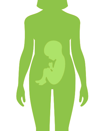
| Foetal anomaly scan This is a mid-pregnancy 20 week scan, detailed scan or anomaly scan is primarily concerned with checking for normal development of your baby’s physical features and internal organs. This scan is perfumed between 18 – 22 weeks gestation .At this stage all of the baby’s organs are developed and clearly visible. Anatomy scan is a very detailed examination where special attention is given to certain organs or features in order to detect any structural abnormalities. In the anomaly scan very detailed checks are made on the flowing anatomical parts of the foetus
|
|
|
|
|
||
| Serial Growth Scans This is called a routine foetal well being or growth scan in which foetal growth and development is assessed through a number of These measurements include
The measurements obtained from the data collected gives an estimate of the foetus weight and possible due date . Doppler of the umbilical artery to assess the blood flow of the foetus from the placenta is performed in every scan.
|
 |
|
|
|
||
| Biophysical Profiling The foetal biophysical profile is used to assess baby’s well-being. This includes a combination of fetal heart rate monitoring (non-stress test) and fetal ultrasound. During the ultrasound the foetal heart rate, breathing, movements, muscle tone and amniotic fluid level are evaluated and given a score. The test is most commonly done when there’s an increased risk of pregnancy loss whereby the obstetrician will determine the necessity and timing of a biophysical profile examination.
|
||
|
|
||
Doppler of major vessels including but not limited to umbilical artery, middle cerebral artery and uterine artery for the assessment of intrauterine growth restriction.
|
||